

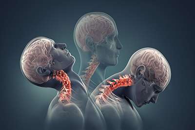
Dynamic MRI Cervical Spine & CCJ Imaging
Establishment of CCJ measurements (Reference Values) for Measures of Craniocervical Stability Using Upright Dynamic Radiography: Summary of Study Findings. (Australia) Reference
Background
The stability of the craniocervical junction (CCJ), which is made up of the occiput, atlas, and axis, is significantly vital. This juncture (joint) permits exceptional motion in the left and right, and flexion and extension. It contains the brainstem, spinal cord, cranial nerves (i.e. nerves in the skull), and other crucial structures. Specialized ligaments and a normal, healthy joint ensure the stability of the CCJ.
Craniocervical instability (CCI) which happens with ligament laxity (an injury/damage to the ligament) or bone abnormalities, can result in an increased range of joint motion that impairs the function of the cranial nerves and central nervous system, and may produce crippling disorders that affect the brain as well as the nerves found throughout the human body and the spinal cord (neurological consequences). CCI has the potential to significantly lower someone’s quality of life as it can result in compression of the neural tissues (affecting the nervous system which regulates and controls body functions) and activity blocks the cerebrospinal fluid (CSF) flow and arterial (blood) supply. CCI can cause a various symptoms ranging from headaches and dizziness to impaired vision and difficulty breathing.
CCI Investigations
Imaging studies are used to help confirm the clinical diagnosis of CCI. Although most imaging methods, such as X-rays and MRI in neutral position, can detect apparent subluxations or neuroanatomical abnormalities, however, they may not be able to clearly show positional abnormalities or minor instability.
Dynamic (also called functional) imaging examinations are advised to help diagnose CCI because symptoms of instability could be more obvious when the head and neck are in dynamic positions, like flexion or extension. Compared to static supine or upright imaging, upright dynamic MRI (udMRI) is a diagnostic method that may assist in the detection of more signs of CCI when the head and neck are in different positions. However, there are a variety of published reference ranges and cutoff criteria, and the radiologist’s reading of craniocervical measurements on udMRI is not uniform.
Study Findings
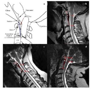
The small study’s findings revealed that most of the four radiological measurements: the basion-axial interval, basion-axial angle (BAA), basion-dens interval (BDI) (FIGURE 1) and the Grabb-Oakes line (GOL)(FIGURE 2) evaluated at neutral, maximal flexion, and maximal extension had significant variations.
FIGURE 1: a) Diagram of Basion-dental interval (BDI) b) MRI-Neutral position c) MRI-flexion d) MRI-extension
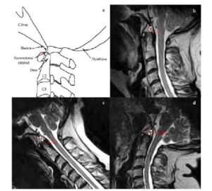
FIGURE 2: a) Diagram of Grabb-Oaks line (GOL) b) MRI-Neutral position c) MRI-flexion d) MRI-extension
Only the BDI and GOL measurements did not exhibit a statistically significant difference when switching from neutral to flexion (TABLE 1).
The findings also revealed that there was no visible difference in any of the three cervical locations between participants who were male and female.
The study also discovered that becoming older was linked to a decline in the parameters in flexion and neutral position only.
Lastly, it was discovered that only the basion-axial interval in the neutral position decreased considerably with aging.
TABLE 1: Reference ranges for radiological measures of craniocervical junction
|
MEASURE |
MEAN |
RANGE (mm) |
|
Basion-axial interval |
Neutral: 4.7 mm Flexion: 5.5 mm Extension: 3.2 mm |
1.0−9.5 mm 0.5–11.0 mm 0.0–8.5 mm |
|
Basion-dens interval |
Neutral: 5.0 mm Flexion: 5.0 mm Extension: 5.6 mm |
3.0–9.0 mm 2.5–11.0 mm 3.0–9.5 mm |
|
Basion-axial angle |
Neutral: 148.6 ° Flexion: 145.8 ° Extension: 161.4 ° |
128.0–168.0 ° 125.0–171.0 ° 128.0–180.0 ° |
|
Grabb–Oakes line |
Neutral: 7.2 mm Flexion: 7.2 mm Extension: 7.2 mm |
4.5–11.0 mm 2. 5–10.5 mm 3.0–10.5 mm |
Note: Standard deviations are not included
Note: XRay imaging is mentioned in this study, however as non-medical professional advice, Cone Beam CT (CBCT) imaging can be a valuable diagnostic tool that provides higher-quality 3D images and more radiographic information compared to XRay images. From our limited understanding, CBCT has significantly less radiation and costs less than a CT Scan. We will keep monitoring for studies regarding this subject however, in the meantime, dynamic CBCT and XRay are excellent means for providing more sensitive measurements compared to MRI; and MRI is an excellent means for providing better information on soft tissues of the craniocervical junction compared to CBCT / XRay.