Atlanto-dental Interval (ADI) – A Radiology Measurement of the Craniocervical Junction
ADI RESEARCH – Several Grade A & B Studies (Excellent!)
Atlanto-dental Interval (ADI) can be affected by numerous diseases, hence multiple research papers look at the changes in the ADI within the patients affected. Last year alone had several grade 1a papers published, and several grade 2a papers. Most newer papers focus on novel treatment methods such as the atlas-profilax technique.
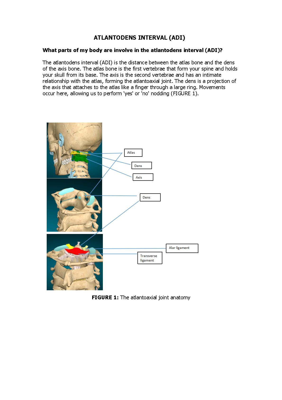
The atlantoaxial junction is the most mobile joint of the body that is held together by ligaments that allow a great degree of freedom of rotation. The atlantoaxial junction is responsible for 50% of all neck rotation, 5° of lateral tilt, and 10° to 20° of flexion/extension. Reference
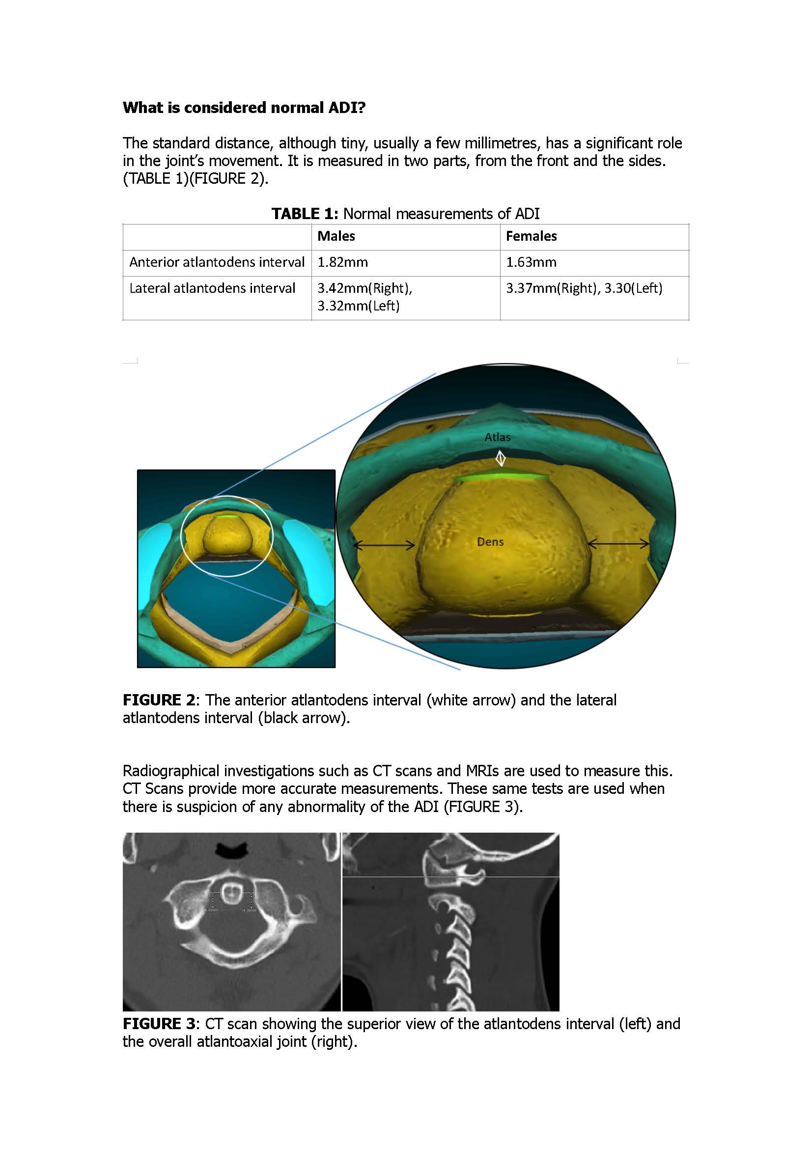
Note: XRays / plain radiography / motion xray is often utilized a the preliminary screening tool; although it is known to be poor quality and therefore less than optimal for measurement accuracy.
What can disturb the normal atlantodental interval?
Any condition affecting the atlantoaxial joint can also affect the atlantodens interval. Common causes include:
- Trauma – One of the most frequent reasons for an irregular ADI is trauma, such as in falls or sports-related mishaps, as this can fracture the dens of the atlas bone.
- Congenital illnesses – Several congenital disorders, including Down syndrome, rheumatoid arthritis, and other connective tissue disorders, can weaken the ligaments holding the dens to the atlas.
- Inflammatory conditions that directly affect the cervical spine.
- Infections
- Tumors that apply pressure on the bones and soft tissues of the cervical spine.
- Iatrogenic causes refer to ADI irregularities caused by medical intervention, such as cervical spine surgeries.
What are some symptoms of an abnormal ADI?
Patients with an abnormality of the ADI, could be experiencing either general or specific symptoms. These are shown in the table below:
TABLE 2: General and specific symptoms of ADI
| General symptoms | Specific symptoms |
| Neck pain | All general symptoms that start from childhood and continue into adulthood can be considered specific to a congenital deformity. |
| Limited range of motion | Feelings of neck instability or a sensation that the neck is not adequately supporting the head following a trauma. |
| Headache | Difficulty in swallowing and speaking is due to compression of the upper spinal cord segment, which controls the function of the pharynx and larynx. |
| Mild neurological symptoms such as numbness in the body | Severe neurological symptoms such as paralysis of limbs, imbalance, and loss of bladder control usually follow spinal cord compression. |
| Muscle spasms | Fever or chronic feeling of illness might be present in the case of osteomyelitis of the atlas or axis. |
What treatment options are available?
Depending upon the severity and cause of the abnormality, the options to treat ADI abnormality are numerous. These include the following:
- General measures include neck immobilization using rigid neck collars.
- Physiotherapy such as training deep and superficial cervical flexors
- In case of any inflammatory disease, non-steroidal anti-inflammation drugs, or in severe cases, steroids can be given to reduce inflammation and allow natural healing.
- In case of infections, antibiotics are given.
- Pain can be controlled by analgesics such as over-the-counter acetaminophen or narcotics. Additionally, muscle relaxant drugs can also be used to treat muscle spasms.
- If the spinal cord is being compressed, surgical intervention is required. Your physician and surgeon will determine the type of procedure following investigations. The operation aims to affix the dens in its usual position and relieve spinal cord compression. Some examples of the methods that could be done include:
-
- Posterior atlantoaxial fusion, in which the atlas and axis are fused with the help of wires and rods to prevent further dislocation.
- Transoral odontoidectomy in which dens is removed by approaching it through the mouth. This is done in case of fracture of dens.
- Anterior odontoid screw fixation involves fixing the dens with the help of screws.
Continue scrolling past references for more important ADI information….
REFERENCES
-
- Chen Y, Zhuang Z, Qi W, Yang H, Chen Z, Wang X, Kong K. A three-dimensional study of the atlantodental interval in a normal Chinese population using reformatted computed tomography. Surgical and radiologic anatomy. 2011 Nov;33:801-6.
- Yang SY, Boniello AJ, Poorman CE, Chang AL, Wang S, Passias PG. A review of the diagnosis and treatment of atlantoaxial dislocations. Global spine journal. 2014 Aug;4(3):197-210.
- Wang C, Yan M, Zhou H, Wang S, Dang G. Atlantoaxial transarticular screw fixation with morselized autograft and without additional internal fixation: technical description and report of 57 cases. Spine. 2007 Mar 15;32(6):643-6.
- Chen Q, Brahimaj BC, Khanna R, Kerolus MG, Tan LA, David BT, Fessler RG. Posterior atlantoaxial fusion: a comprehensive review of surgical techniques and relevant vascular anomalies. Journal of Spine Surgery. 2020 Mar;6(1):164.
- Elbadrawi AM, Elkhateeb TM. Transoral approach for odontoidectomy efficacy and safety. HSS Journal®. 2017 Oct;13(3):276-81.
- Munakomi S, Tamrakar K, Chaudhary PK, Bhattarai B. Anterior single odontoid screw placement for type II odontoid fractures: our modified surgical technique and initial results in a cohort study of 15 patients. F1000Research. 2016 Nov 21;5(1681):1681.
- Michel C, Dijanic C, Abdelmalek G, Sudah S, Kerrigan D, Yalamanchili P. Upper cervical spine instability systematic review: a bibliometric analysis of the 100 most influential publications. Journal of Spine Surgery. 2022 Jun;8(2):266.
- Joaquim AF, Appenzeller S. Cervical spine involvement in rheumatoid arthritis—a systematic review. Autoimmunity reviews. 2014 Dec 1;13(12):1195-202.
- Shah A, Vutha R, Prasad A, Goel A. Central or axial atlantoaxial dislocation and craniovertebral junction alterations: a review of 393 patients treated over 12 years. Neurosurgical Focus. 2023 Mar 1;54(3):E13.
- Manent L, Higuera JG, Lewis K, Angulo O, Solano ME. Radiological Improvements in Symmetry of the Lateral Atlantodental Interval and in C1 Tilt After the Application of the Atlasprofilax Method. A Case Series. Qeios. 2022 May 20.
What other ADI information might be helpful?
“Traumatic flexion of the neck may result in injury to the transverse odontoid ligaments and alar ligaments. An atlanto-dental (i.e. C1-C2) interval over 3 mm suggests possible instability in adults; an ADI exceeding 5 mm suggests instability in children.
An ADI of 7 mm suggests rupture or incompetence of the transverse ligament, and/or of the cruciate ligament, and an ADI of 10 mm suggests loss or incompetence of the alar ligaments as well. However, if the transverse ligament is intact, the ADI is normal despite the presence of alar ligament incompetence.
A proclivity to ligamentous incompetence renders the atlantoaxial joint at higher risk for instability. The atlantoaxial junction is the most mobile joint of the body that is held together by ligaments that allow a great degree of freedom of rotation, the atlantoaxial junction is responsible for 50% of all neck rotation, 5° of lateral tilt, and 10° to 20° of flexion/extension…” Reference
Atlanto-dental Interval (ADI) is utilized to help diagnose Atlantoaxial Instability (AAI)
Atlantoaxial instability (AAI) is characterized by excessive movement at the junction between the atlas (C1) and axis (C2) as a result of either a bony or a ligamentous abnormality/injury. [1]. The normal range of motion for rotation at the joint is 40 degrees. The excess mobility at the articulation can lead to AAI.
Atlantoaxial instability can originate from congenital conditions, but in adults, it is primarily seen in the setting of acute trauma or degenerative changes due to the inflammatory response of rheumatoid arthritis (RA).
Infection has been found to be an additional cause of instability, with the rich arterial supply and venous plexus in this region of the body providing a route for infectious sequelae…” Reference
AAI can also occur in connective tissue disorders [4].
Dynamic Imaging for a Comprehensive Assessment
The stability of the craniocervical junction (CCJ), which is made up of the occiput (base of skull), atlas (C1), and axis (C2), is significantly vital. This juncture / joints permit exceptional motion in the left and right, and flexion and extension. It contains the brainstem, spinal cord, cranial nerves (i.e. nerves in the skull), and other crucial structures. Specialized ligaments and a normal, healthy joint ensure the stability of the CCJ.
Dynamic (also called functional) imaging examinations are strongly advised to help diagnose atlantoaxial instability because symptoms of instability could be more obvious when the head and neck are in dynamic positions, like flexion or extension.
This means that the radiology technologist will take the diagnostic radiology images in neutral, flexion and extension and the radiologist should measure Atlanto-dental Interval (ADI) in neutral, flexion and extension, although this rarely seems to occur right now because dynamic imaging is a relatively new modality and most physicians are not up to date on best practices, in fact, some may argue that flexion and extension positions are unnecessary. Spine and Brain Advocate’s Consultant Radiology 2nd Opinion services can provide you with a comprehensive, detailed expert report.
Radiology Measurements
There are several key radiology measurements that are universally accepted by majority of neurosurgeons around the world and Atlanto-dental Interval (ADI) is one of them. To learn more about some other craniocervical junction radiology measurements, you can visit our Radiology Measurement page.
Other Important Considerations
Since the first two cervical vertebrae, the atlas (C1) and the axis (C2), contain vital neural and vascular structures (i.e. brainstem, spinal cord, cranial nerves, vertebral arteries and internal jugular veins, it may also be important to consider vertical craniocervical instability, intracranial hypertension / hydrocephalus as well as cerebrospinal fluid flow and internal jugular vein blood (IJV) flow issues due to elongated styloid processes (Eagles Syndrome) or atlas (C1) impingement.
SBA Report Outcomes
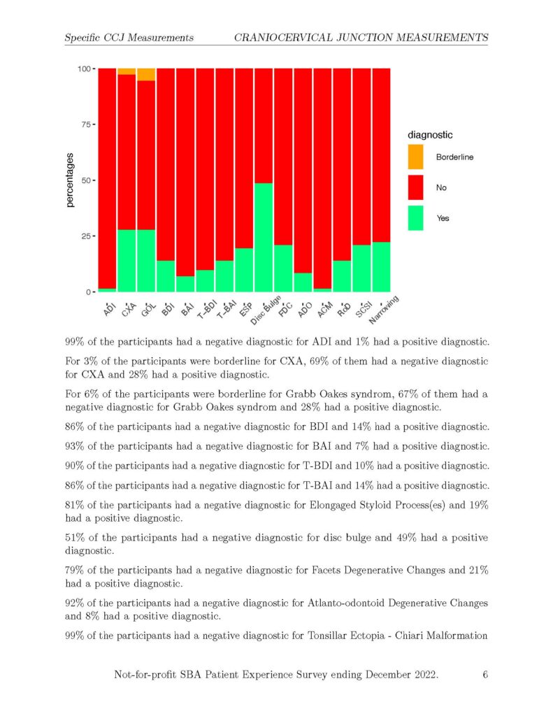
In SBA’s Radiology 2nd Opinion Report Results ending December 2022, only 1% of the participants had a positive abnormal ADI measurement (see image below). To read more about SBA’s Radiology 2nd Opinion Report Summary visit here.
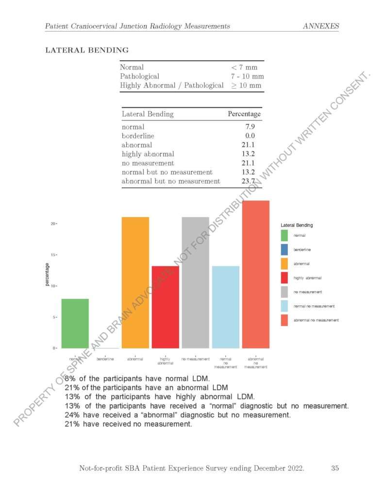
 In SBA’s CCJ Patient Experience Survey Results ending December 2022, there was no information gathered on ADI, however Lateral Mass Displacement (LMD) information was presented on page 35. While LMD is not the same as ADI, both measurements can help diagnose atlantoaxial instability. Lateral Mass Displacement refers to the displacement of the C1 lateral masses on C2. This is sometimes called Lateral Mass Overhang or Rule of Spence.
In the CCJ Patient Experience Survey Results ending December 2022, 21% of the participants had an abnormal LMD measurement (7mm – 9.9mm LMD) and 13% of the participants had a highly abnormal LMD measurement (equal or greater than 10mm).
Haven’t completed or submitted an updated CCJ Patient Survey? You can help lobby for healthcare changes by completing the CCJ Patient Experience Survey here.
Shop 2nd Radiology Opinion Services and Products
Important: Having access to your medical information is a powerful thing and an important part of advocating for your healthcare. However, this website should never replace advice from your physician who understands your specific medical history and is trained to care for patients.
If you have any suggestions or see any errors on this page, please contact us.
SBA Thanks Professor of Medical Education, Dr. A. Bohari for contributing to this page.
