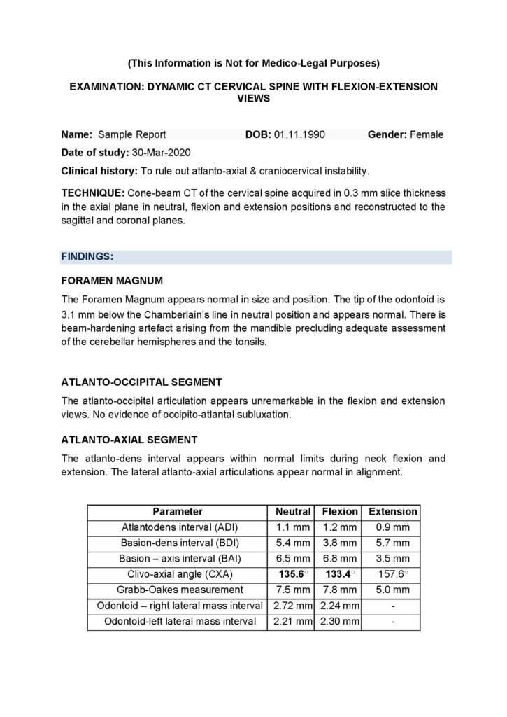
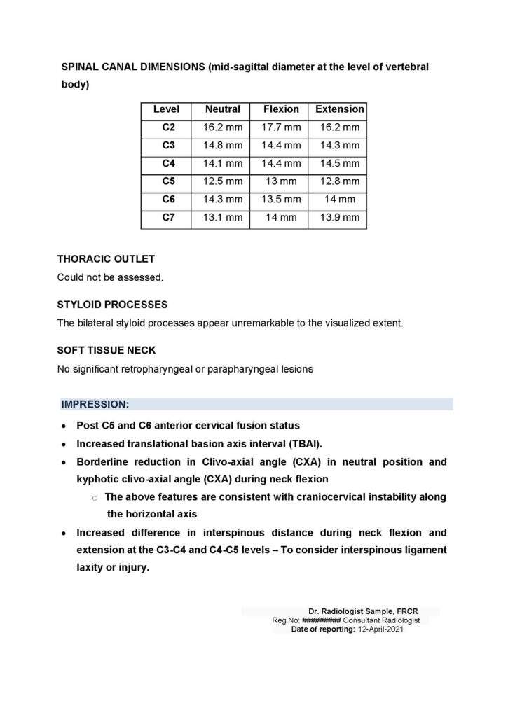
Below is a sample report of a Dynamic CT Cervical Spine and Craniocervical Junction in neutral, flexion and extension positions. This report demonstrates the level of detail and thoroughness of our radiology reports.
It is important to keep in mind that the reports written by certified radiologists, significantly depends on quality of the study images provided such as the sequences obtained, the visual quality of the images, and the technical aspects of the imaging. Ideally, CT scans or upright Cone Beam CT scans, should be completed in less than 1 mm slices to render 3D images for further assessment of key anatomy such as the joints between the first two vertebrae (C1-C2) and the base of skull and C1, and the area of the first two vertebrae and other bony structures such as the styloid processes that can assist in examining if vascular compression by bones may be a factor for further assessment.
To learn more about today’s gold standard imaging details, please refer to Book 1 in a 3-book series: A Patient’s Guide To Craniocervical Instability Diagnosis


